
Three-level validation.
To reduce uncertainty.
Partners in Excellence
Preferential conditions for beneficiaries.
Please check your policy with your insurer or contact us for further clarification.
Not one reading. Three.

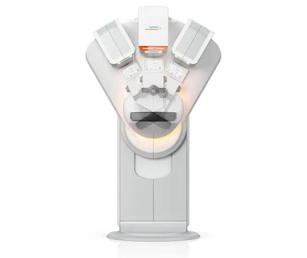
High precision mammography.
AI based image analysis.
Specialized medical review.


WHAT TO EXPECT
Your visit, step by step
From arrival to your report, every stage of your visit is carefully guided.
Arrival and registration
On arrival, our concierge welcomes you and accompanies you throughout the process.
The environment is designed to support comfort, privacy and calm.

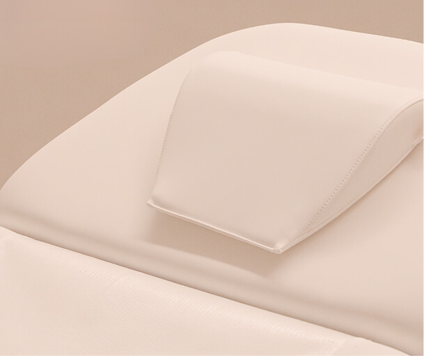
Preparation and Guidance
The technician accompanies you throughout the entire individual process, ensuring your comfort and privacy. She explains the procedure, answers all your questions and guides you through correct positioning.
Private environment and curated support.
3D Image Acquisition
The equipment captures dozens of images from different angles, reconstructing the breast in three dimensions. Artificial intelligence analyses each layer simultaneously, identifying areas that may require attention.
Low radiation technology, within established safety limits.
High-Definition
Breast Ultrasound
After the mammogram, you are immediately guided to the ultrasound room, where the radiologist complements the 3D images with ultrasound, allowing a comprehensive breast assessment.
Integrated pathway with no waiting time.
Report Issuance
The radiologist analyses all the information collected during the exam in order to issue the report: the 3D images, the breast ultrasound and your clinical history.
Careful analysis of all clinical information.


FREQUENTLY ASKED QUESTIONS
Questions and Answers
Does the exam take longer than a traditional mammogram?
No. Image acquisition time is very similar, approximately 7 seconds per view. The image acquisition takes 5 to 7 minutes.
Is radiation exposure higher with 3D mammography?
No. The equipment used performs 3D mammography with a low radiation dose, keeping exposure within internationally established safety limits. Radiation levels are equivalent to or lower than those of standard 2D mammography.
Can I have the exam if I have breast implants or surgical scars?
Yes. 3D mammography can be performed in women with breast implants. Surgical scars, fibrotic tissue or previous post surgical changes do not prevent the examination. Please inform the technical team before the exam about any previous surgery to allow optimal positioning.
When will I receive my results?
The medical report is issued within five working days after examination. An expedited option is available upon request. If any findings require prompt attention, you will be contacted immediately by our clinical team.
Why are images acquired from different angles?
During tomosynthesis, the X ray tube moves in an arc over the breast, capturing 15 to 25 images from different angles. Each image corresponds to a thin layer of breast tissue, allowing a full 3D reconstruction and improving the detection of very small lesions that may not be visible on a single flat image.
IMPORTANT INFORMATION
3D mammography is an imaging exam that complements clinical breast assessment and does not replace a medical consultation. All images must be interpreted by a qualified expert. Findings may require additional imaging and or further tests. A normal result does not completely rule out disease. If you notice symptoms, changes, or a lump, please seek medical advice regardless of your results.